Prof. Nicolas Baeyens
Université Libre de Bruxelles
Could you please tell us about your research field?
I am a cardiovascular physiologist specializing in tissue mechanical homeostasis, in other words, how cells integrate mechanical forces to control tissue remodeling and architecture.
For example, I am interested in abnormal vascular remodeling events such as arteriovenous malformations or aneurysms and cardiac fibrosis, an abnormal scarification process with deadly consequences. I am also focused on the composition of the extra-cellular matrix (e.g. collagen, fibronectin proteins) and its modulation during the time that impacts how cells behave.
To note, blood itself can send a physical signal to endothelial cells because it flows on them having frictional force, fluid shear stress. I spent most of my post-doc investigating how cells sense shear stress, integrate it, and communicate according to the force perceived. In other words, if the force is increasing or decreasing, blood vessels remodel thanks to a cell signaling cascade promoting this process.
What is challenging in the mechanical force research field is that there are so many parameters in each stimulus, many physiological variables to consider (e.g. flow direction, intensity, frequency), leading the combination of different stimuli and their receptors to be very complex.
In more details, what are you looking for inside a biological sample and which are your microscopy requirements?
When I look for cells responses inside a three-dimensional organization, I need to know very precisely how the force distribute. In addition to that, I need to get the sense if the vessels are normal or not, if cells are in shape or stretched, if the matrix has been impacted somehow.
“I need precise information both at single-cell level but also in the neighboring environment”
This scientific interest leads to a microscopy approach with a large field of view to appreciate what is happening in the whole sample. I work with tissues, with organoids, with cells organized in 3D where I have to apply forces on top of them, and for this aim, the ideal configuration is an up-right microscope. Besides that, the objective choice is also crucial since the samples I want to image are often thick and very large. As I investigate the impact of forces generated by a dynamic and fast blood flow, I need to perform fast imaging to perform tracking analysis and fluid displacement measurements to map fluid shear stress accurately.
Can you please describe your up-right microscope set-up and the different biological applications that you could develop with it?
The up-right microscope set up is organized around a Nikon FN1 microscope with a FTP-2000 stage equipped with a 300 um piezo plate (ASI). The idea behind was to allow enough room for “unorthodox” samples to be imaged. On top of that we chose to go with the Celesta laser source (Lumencor) and a Prime BSI camera (Photometrics). We therefore get 18 mm field of view with 7 different wavelengths and the capability to image at up to 1 kHz. I mainly use water immersion objectives.

Thanks to the up-right architecture I build up in my lab, I can play with different applications:
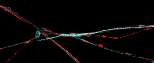
- imaging red blood cells displacement and vessel remodeling in Zebrafish larvae
- imaging fast fluorescent beads displacement in millifluidics channels
- imaging fast cellular calcium responses
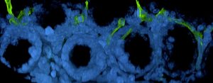
- imaging of transparized tissue with dedicated multi immersion objectives (in collaboration with Nikon).
In my lab, many students use the microscope with different samples, with vastly different acquisition parameters, on the same day. I have to say that the combination of the CrestOptics system with the NIS Elements software (Nikon) works very well all of them, allowing for smooth transition between each experiment.
“The microscope equipped with X-Light V3 represents a very versatile platform, and it is adaptable with excellent results to the multiple applications I work on”
Could you please tell us which features of X-Light V3 confocal spinning disk have helped your research?
As I mentioned before, I much appreciate the high speed and the large field of view of the X-Light V3, compared to the other microscopes that I used before. Moreover, a nice feature of the CrestOptics system is that just with a “click” I can switch between widefield and spinning disk mode and disk replacement is very simple. I also appreciate the reliability of the system, allowing me to reproduce the data easily. All in all, the system has many excellent features, and at the same time, it is affordable.
“At the end of the day, you not only need a nice picture to show but also take out data and graph from it, and that is what I can do with X-Light V3”
When I started with transparized 3D tissue, I was planning to use a light-sheet microscope, but as soon as I tried your spinning disk, I realized that I could ideally use the V3 for that application with a successful outcome. So far, we optimized various clarification and staining methodologies to check the whole vascular network in health and pathological tissues with a thickness of several millimeters and get both quantification and beautiful views with the X-Light V3.
“Thanks to the combination of transparized tissue and an excellent microscope, we can do things that, honestly, I could not imagine doing a few months ago!”
How do you see the evolution of fluorescence microscopy to keep up with innovative biological applications?
We need to image large volumes at high resolution with multiplexing, primarily to investigate structure/function relationships in health and disease conditions. This need requires fast and precise acquisition, so I guess microscopy has to keep developing in that direction.
In particular, for my research, I would also love to have the same system on an inverted architecture with silicone oil objectives to go reach subcellular resolution in relatively thick samples.