Dr. Sylvie Le Guyader
Live Cell Imaging Facility Manager, Karolinska Institutet, Stockholm
Dr. Gabriela Imreh
Microscopy Engineer, Karolinska Institutet, Stockholm
Please, tell us about your experience in microscopy and your roles at the Live Cell Imaging facility at the Karolinska Institute.
Sylvie Le Guyader
“I have a background as a biologist. I got a PhD and continued as Post Doc in different Institutes having the chance to collect lot of experience in microscopy. I also gained experience in helping people buy microscopy equipment which led me to creating the Live Cell Imaging facility at the Karolinska Institute. I have been the manager of the LCI Facility for 13 years.”
Gabriela Imreh
“At the LCI facility, I am in charge of training people on the different microscope set-ups we have. In our facility, we offer very extensive trainings on the instruments to make sure the users become fully autonomous in image acquisition and image analysis for their own samples. We are also always there after training if they need help or expert advice. For us, it is key that the users fully understand what they are doing so they can choose which objective and settings work best for their scientific question. During the training, they get the chance to test different settings so they fully understand the imaging outcomes.
“The aim for us is that our users become independent microscopists, able to select the right microscope settings based on their sample and scientific question.”
In a facility you have the chance to deal with many types of microscope set-ups. In your experience, which are the main advantages of CrestOptics X-Light V3?
We bought the CrestOptics X-Light V3 spinning disk about one year ago and we have a lot of experience with it. We bought it after having already experienced other spinning disk systems with some limited features in terms of field of view, speed and resolution. So, we needed to have an upgraded spinning disk. CrestOptics X-Light V3 is a great update thanks to the large field of view and to the fact that it is designed for two cameras. Our choice is to have a Prime BSI camera and a 95B camera (Photometrics) with different pixel size to satisfy Nyquist sampling rate with a range of objectives with different magnifications.
“The facility users are very happy with CrestOptics X-Light V3 spinning disk.”
The other relevant feature our users appreciate is the possibility to have an easy bypass mode from Widefield to Spinning disk illumination. The users are trained to acquire tiled images of their sample in widefield with maximum speed and minimum illumination. They then use the tiled image to navigate in the sample and decide where they want to go without further bleaching the sample. They can then move to the area of interest and switch to Spinning Disk mode to acquire confocal images.
“Facility users appreciate a lot the easy switch from Widefield to Spinning disk modality, giving them the possibility to select the proper imaging according to their application and experimental needs.”
Could you please describe in more details the microscope set-up equipped with CrestOptics X-Light V3 and for which applications is mainly used?
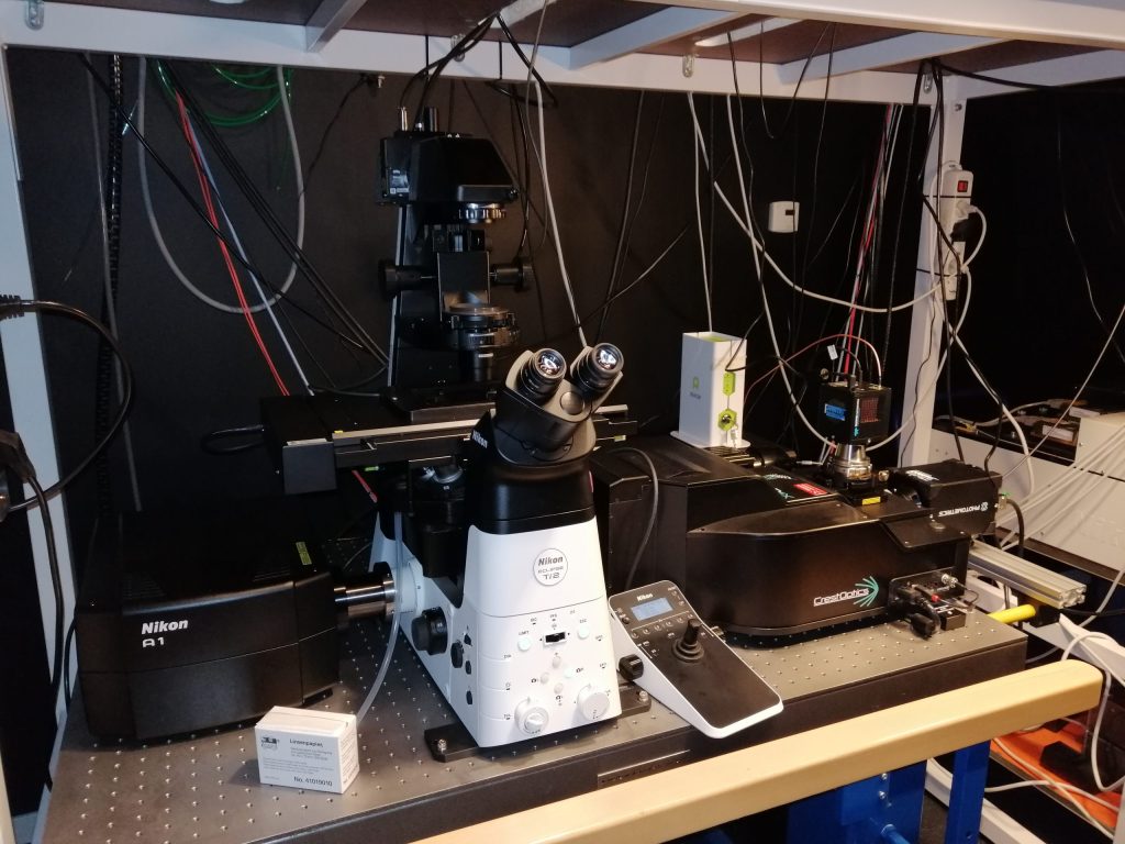
Our system consists of a Nikon Ti2 equipped with X-Light V3 spinning disk on one port and a single point confocal on the other port. This allows us to combine different imaging modalities. Recently, we upgraded the system with a CO2- and temperature-controlled incubator to enable imaging live samples.
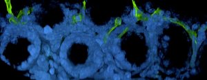
Concerning biological applications, we image a wide variety of samples on the CrestOptics spinning disk. The most common ones are monolayer of cells stained for different protein markers, tissue sections of different origins and thickness as well as organoids.
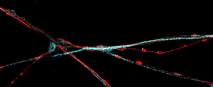
Currently, we are working on implementing FRAP (Fluorescence Recovery After Photobleaching) by bleaching with the single point confocal followed by fast imaging on the spinning disk confocal side. The FRAP method is widely used to study protein mobility in living cells by photobleaching a specific area of a cell by intense laser light, in other words removing fluorescence from this area, and hence quickly monitoring the fluorescence diffusion throughout the sample until the replacement of non-fluorescent probes in the bleached region
“The users we train on the X-Light V3 are those who need to image fast and with a large field of view but still need optical sectioning.”