Please, tell us about your scientific background and your experience in the field of microscopy.
I have a PhD in biophysics and molecular cell biology, and I have been doing science for more than 20 years. I have worked most of my academic life with custom-made microscopes and I started to work with commercial instruments recently. I think it is easier to teach someone when you have a commercial instrument; using custom-built microscopes you have to spend days before you start to feel comfortable using the machine.
“I think it is easier to teach someone when you have a commercial instrument”.
How is the European Molecular Biology Laboratory (EMBL) organized and how do you help research groups with your facility?
EMBL is a pan-European Life Science institution. It is the major molecular biology life science institute with several sites across Europe, such as Hinxton (England), Barcelona (Spain), Heidelberg (Germany), Rome (Italy), Hamburg (Germany) and Grenoble (France).
The Microscopy Facility provides users with access to state-of-the-art instrumentation as well as with training, image processing and quantitative image analysis. The big part of our facility’s mission is to provide services and expertise to all the internal EMBL members as well as for external users.
"The big part of our facility’s mission is to provide services and expertise to all the internal EMBL members as well as for external users. "
Which systems are present in your facility and why did you choose the CrestOptics X-Light V3 spinning disk confocal? And how did you find the use?
When I started here, in 2019, we had two scanning and two spinning disk confocal microscopes. I used a lot the scanning confocal system for spectroscopy but, I have to confess, I don’t like it for imaging (why do you have to spend an hour for a single field of view Z stack?). Meanwhile, the spinning disks in our facility were starting to become obsolete, so I began to consider replacing them. I found the CrestOptics X-Light V3 spinning disk with the possibility to have two cameras and larger FOV, so we needed all these improvements with respect to our previously generation spinning disks. Moreover, I liked the fact that CrestOptics is a local company and wherever you are dealing with an innovative company is good if you have it around, this can make life easier. The CrestOptics X-Light V3 spinning disk had all the potentiality that I needed: simultaneous fast dual-color imaging, a wider FOV and I also liked the fact that the spinning disk is enclosed on a vacuum seal chamber, this avoid many problems.
In addition, the cost, the modularity, the features mentioned above made the CrestOptics X-Light V3 spinning disk the right instrument to choose.
In particular, moving to the V3 from scanning confocal microscopy we appreciated the speed; for example, with the laser scanning it took 40 minutes or more to make a Z-stack, now only 3 minutes and it was a massive improvement.
The use of the spinning disk is fantastic, you can acquire even more data and in a shorter time; for most common imaging the CrestOptics X-Light V3 spinning disk is perfect, and it became our default imaging system.
“The use of the spinning disk is fantastic, you can acquire even more data and in a shorter time; for most common imaging the CrestOptics X-Light spinning disk is perfect, and it became our default imaging system”
Could you please describe your microscope set-up equipped with CrestOptics X-Light V3? How do your users respond to its use?
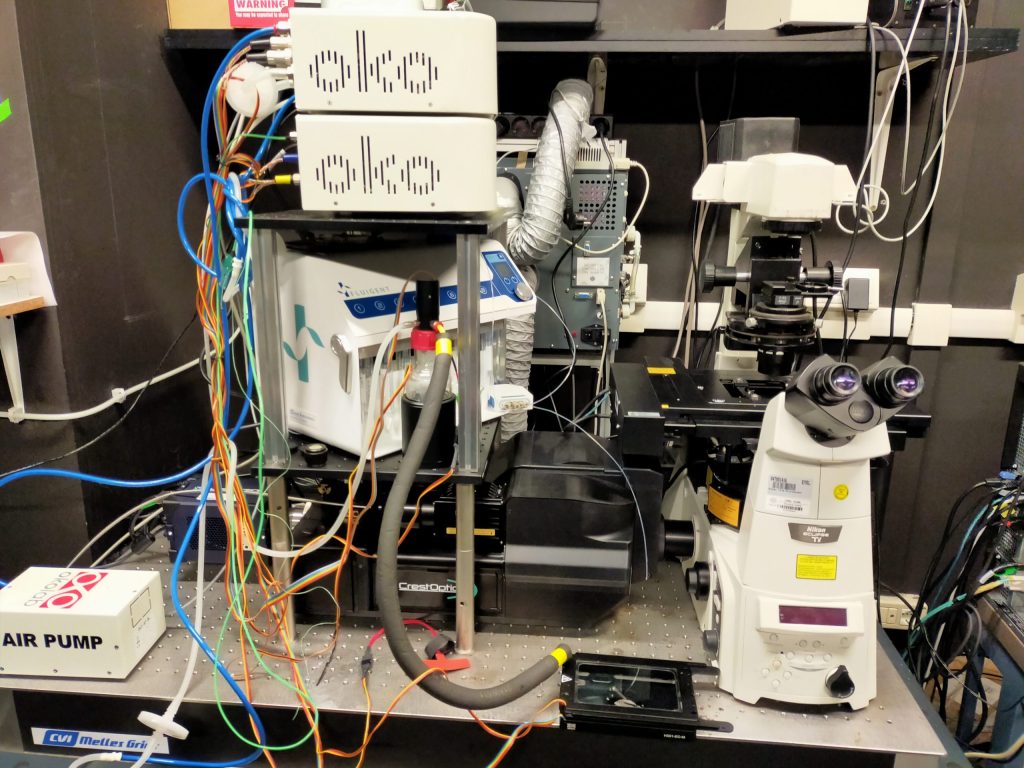
At the moment, we use the CrestOptics X-Light V3 spinning disk with a Nikon Ti inverted microscope, a Prime BSI camera and a Celesta laser source. We coupled this system with fluidics; in particular, we are doing an exchange experiment, like seqFISH+, a particular method for spatial transcriptomics. It was straightforward to integrate the fluidics system on the microscope because NIS Elements software can easily read the TTL triggers via a simple USB National Instruments card. We tried with other microscope brands but it was a nightmare.
You have had the opportunity to use the DeepSIM in your facility, what are your first impressions and what advantages would it bring to your microscopy lab?
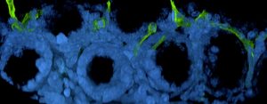
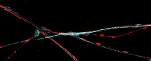
I had the opportunity to test the DeepSIM in an alpha version and it was good. I had also another lattice SIM-based super-resolution instrument for almost one year in my facility and in general, for tissues, it was not amazing. In terms of flat samples, the DeepSIM performed similar to the other super-resolution system, but with the DeepSIM it is possible to go deep through tissues and the reconstruction in this kind of samples was very good. This is very important for us since 90% of our samples are tissues and we want to get the best for it; that also means that we don’t only want to use high magnification objectives, but we want to have super-resolution also with 20X and this is only possible with the DeepSIM.
Therefore, DeepSIM works also with low magnification objectives and applying this feature for tissue and expanded samples imaging is very useful. Obtaining super-resolved images also with low magnification objectives is a unique feature of DeepSIM and it is very important for tissue imaging. Moreover, the DeepSIM is taking the widefield to the next level because at the end you have a widefield illumination, so you need a little amount of light but with the improvement of super-resolution and that’s really cool in terms of live imaging.
“We want to have super-resolution also with 20X and this is only possible with the DeepSIM”
How important is to have a super-resolution instrument in a modern microscopy facility and what are the DeepSIM features that most impressed you?
It’s a must for a microscopy facility; if one of our mandates is to offer state-of-the-art infrastructure and instrumentation, we need to be able to provide super-resolution.
I appreciated the DeepSIM ability to perform deep imaging and also the possibility to do super-resolution with low magnification objective.
“It is a must for a microscopy facility; I appreciated the DeepSIM ability to perform deep imaging and the possibility to do super-resolution with low magnification objective”
How do you see the evolution of fluorescence microscopy to keep up with innovative biological applications?
Probably one of the most important things to come is SMART microscopy, the fact that you would automate experiments through the use of artificial intelligence, machine learning or deep learning and this would impact a lot how you do experiments. It’s all about automating the experiments and acquisitions, this is one of the major things that will happen in the next 5 years.
“It’s all about automating the experiments and acquisitions, this is one of the major things that will happen in the next 5 years”