Brain organoid: visualization of cortical neuron subtypes

基于当前为阻止SARS-CoV-2新型冠状病毒传播而实行的限制策略,我们在意大利科技学会(IIT)- CLNS SAPIENZA的合作者与我们分享了其样品:厚度为50 μm的大脑类器官切片。我们用CrestOptics X-Light V3 转盘式共聚焦系统获取了图像。

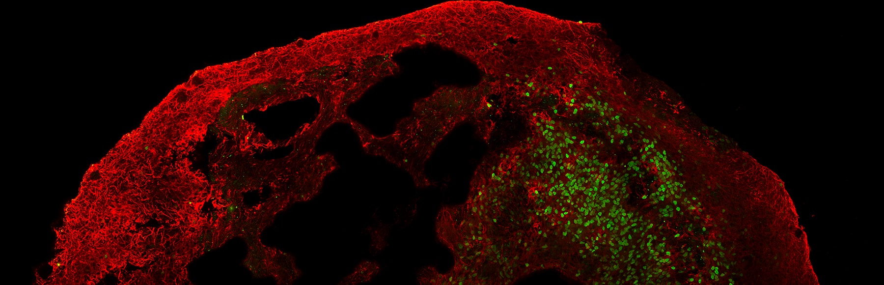
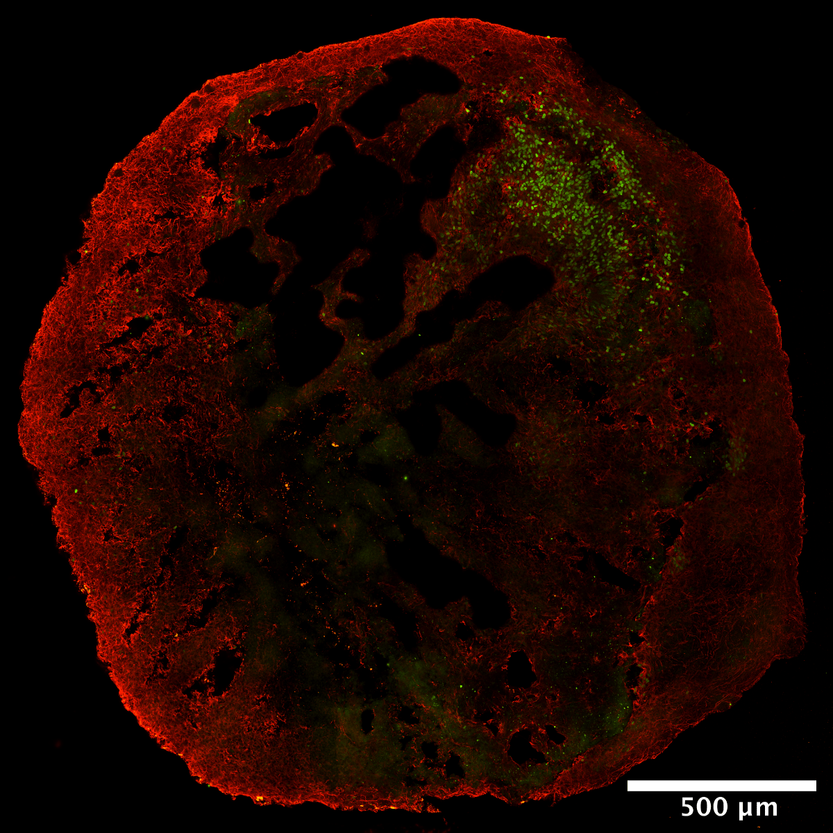
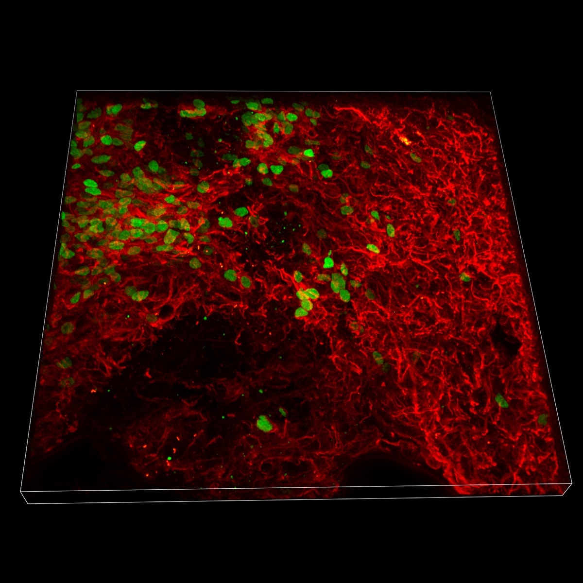
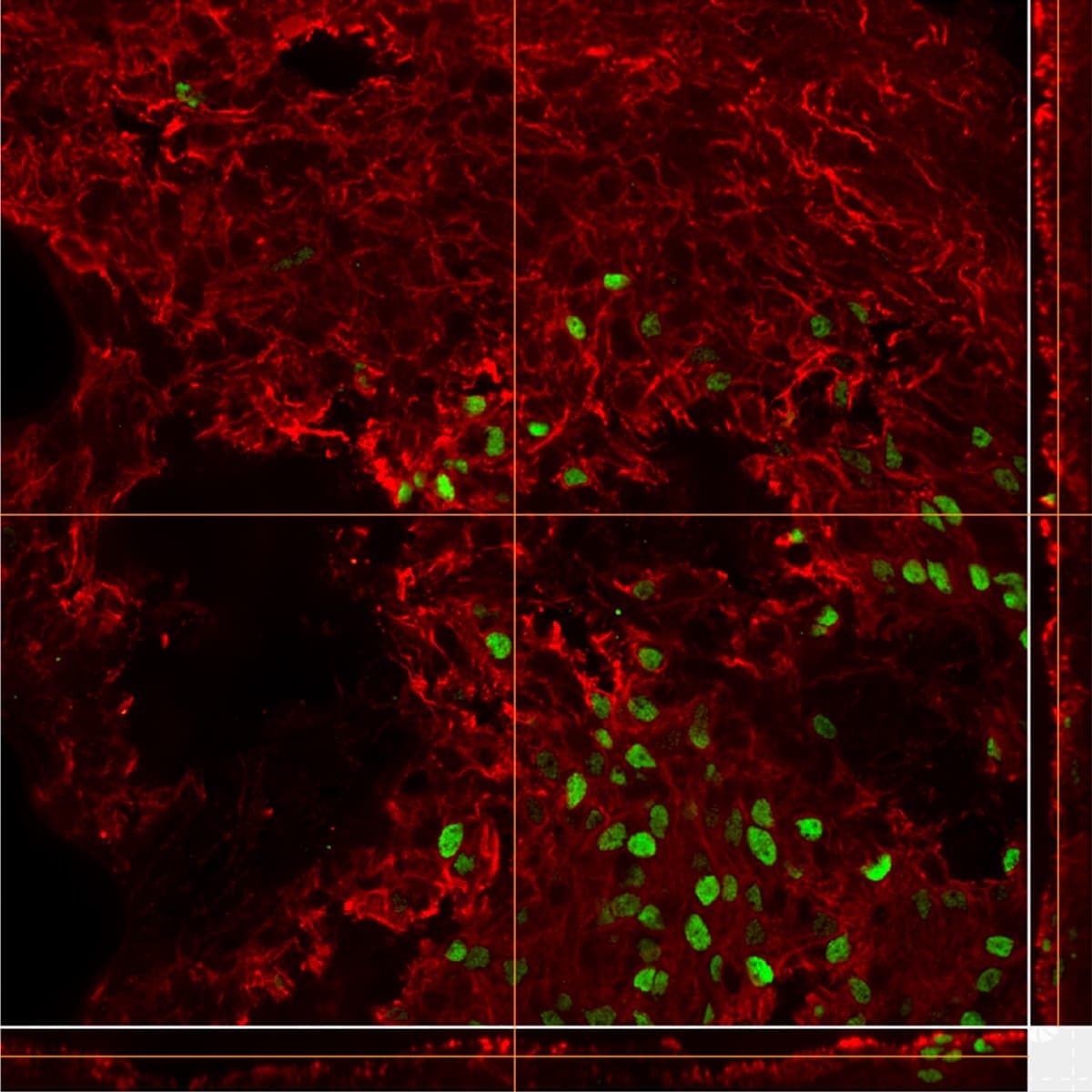
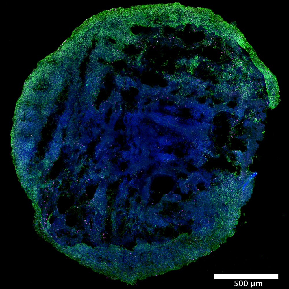
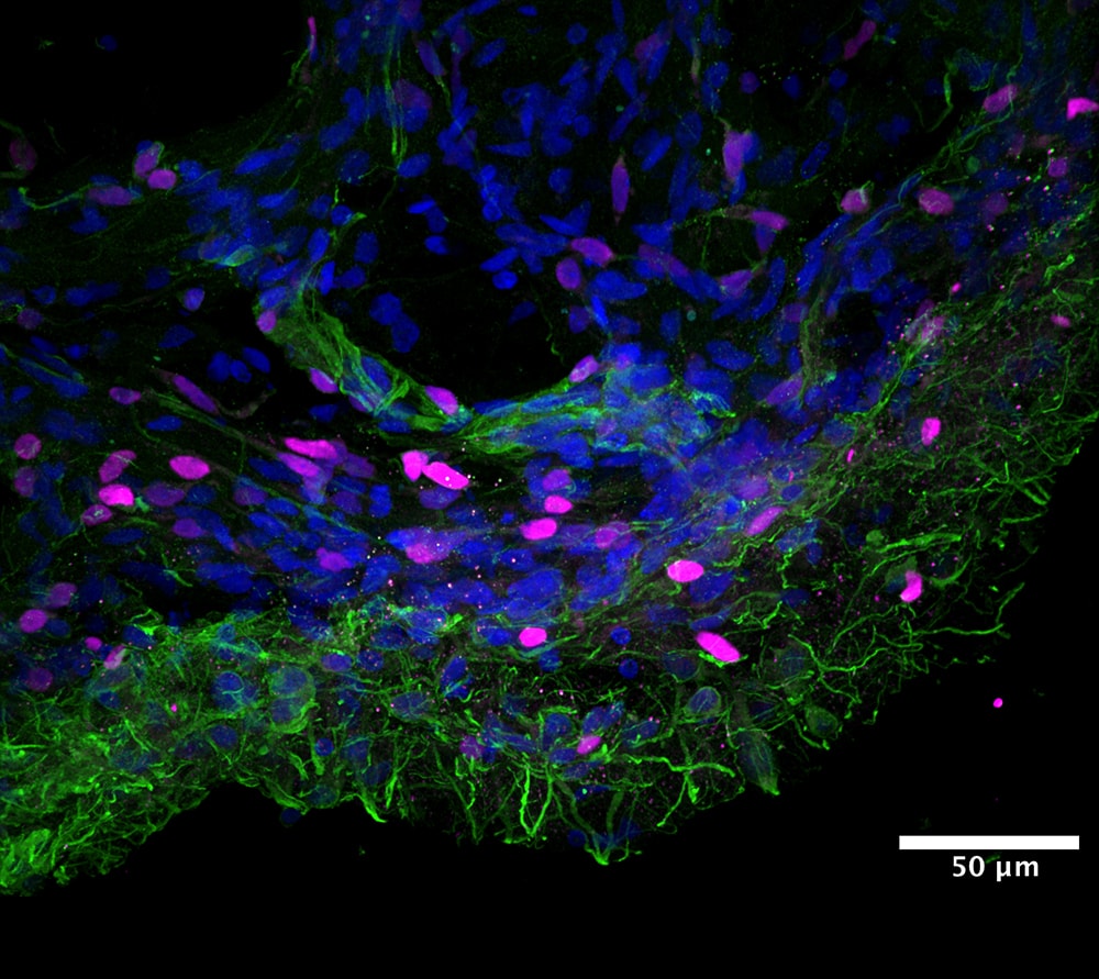
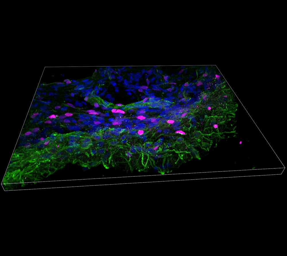
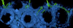
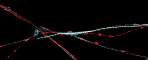
In particular, brain organoid structures shown here derive from in-house derived control iPSC line grown for 71 days under continuous shaking on a 3D orbital shaker in order to ensure the homogeneous diffusion of the media nutrients within the structures. Then, organoids were fixed, sliced and sections were stained for various differentiation markers (described in Figure A and Figure B) in order to confirm the generation of specific cortical neuron subtypes.

Figure A: cortical neuron subtypes

  • MAP2 (Alexa 594 fluorophore, shown in red), a microtubule-associated protein present in the cytoskeleton of neuronal dendrites.
  • CTIP2 (Alexa 488 fluorophore, shown in green), which stains cortical neuron nuclei that localize in the layer V of the cortical plate and its expression is involved in the early specification of a distinct class of cerebral projection neurons.

拼接图像

(Plan Apochromat Lambda 60X oil, 1.45 NA)

3D 体积重建及正交(XZ、YZ平面)视图

(Plan Apochromat Lambda 60X oil, 1.45 NA)

Figure B: cortical neuron subtypes

  • TBR1 (Alexa 750 fluorophore, shown in magenta) a nuclear transcription factor important for the glutamatergic neuron differentiation during the cortical development and its staining is specific for those neurons that localize in layer VI of the cortical plate.
  • MAP2 (Alexa 488 fluorophore, shown in green), a microtubule-associated protein present in the cytoskeleton of neuronal dendrites.

Stitched image

(Plan Apochromat Lambda 60X oil, 1.45 NA)

最大强度投影及 3D 体积重建

(Plan Apochromat Lambda 60X oil, 1.45 NA)

Share this Article

Fill the form and download the Application Note

Fill the form and download the Application Note