
Prof. David J. Hodson
Institute of Metabolism and Systems Research (IMSR), University of Birmingham, Birmingham, UK.
Centre of Membrane Proteins and Receptors (COMPARE), University of Birmingham and University of Nottingham, Midlands, UK.
Centre for Endocrinology, Diabetes and Metabolism, Birmingham Health Partners, Birmingham, UK
Could you please tell us about your research field?
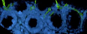
I am mainly focused on pancreatic islet biology. The pancreatic islets are responsible for glucose homeostasis through the production of hormones such as insulin, glucagon and somatostatin. During type 1 and type 2 diabetes the islet produces insufficient or inappropriate levels of hormone, leading to elevated (or too low) blood glucose levels, which drives a range of complications (kidney and heart issues, nerve and vascular problems, eye problems, delayed wound healing). Together, type 1 and type 2 diabetes affect ~ 10% of the UK population and as such are significant healthcare issues.
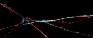
Together with collaborators, we are also interested in technology development, particularly in optogenetics, chemogenetics, opto-chemical and enzyme self-labeling approaches. For instance, I am involved in the development of next generation snap-tag fluorescent labels in collaboration with a numbers of researchers across Europe and US.
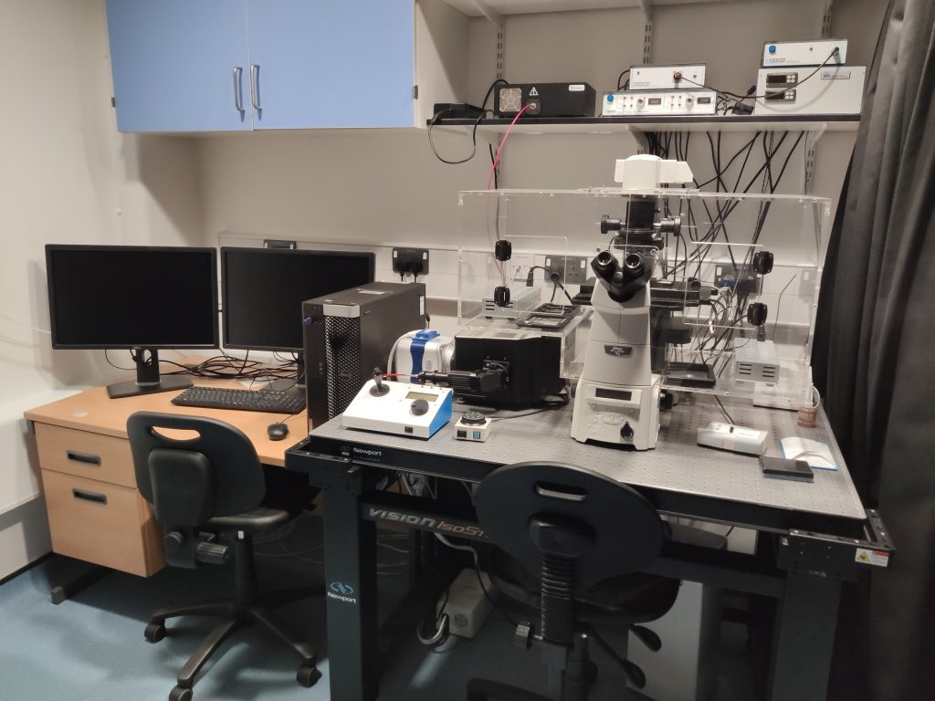
When you had to buy a new microscope, why did you choose CrestOptics X-Light spinning disk?
I always had a long-standing relationship with Cairn Research, a company specialized in microscope solutions for the life sciences and they, as CrestOptics distributor, recommended to me the CrestOptics X-Light confocal spinning disk. I immediately took the recommendation seriously since I was looking for a flexible system suitable also for fast live-imaging, for example for the screening of calcium, cAMP and ATP/ADP dynamics using fluorescent indicators or genetically encoded probes like FRET probes.
“X-Light spinning disk has the ability to record very quickly and detect dynamic events over long periods of time. It allows us to capture and register the fast cellular changes we would otherwise miss”
Another strong point of X-light spinning disk is the flexibility in terms of camera and light source combination, meaning that the system can be easily upgraded and does not become obsolete like others. In my case, I started with a epifluorescence and an EMCCD-based configuration, but have recently upgraded to a multiline laser to achieve more power at the sample for confocal microscopy and single molecule FISH.
How X-Light spinning disk has helped your research?
In my group, we have used X-Light spinning disk in most of our published studies and there are lots of ongoing collaborations involving the system. It is exceptional for dynamic live-imaging of cells and is supremely flexible in terms of software and hardware capability.
“The X-light V2 spinning disk enables us to routinely perform complicated live-imaging experiments for extended periods of time, using a variety of probes spanning dyes through genetically-encoded probes”
Can you mention other biological applications which were developed after the system has been installed in your lab?
We have a numbers of collaborators in the US who we do a lot of imaging for. I would like to mention Prof. Franck Mauais-Jarvis at Tulane, New Orleans. His group studies sex differences in pancreatic islet function and we do lots of cyclic AMP imaging with him.
Regarding development of chemical probes, we closely collaborate with Dr. Johannes Broichhagen at Leibniz FMP in Berlin with whom we create fluorescent labels for conventional and super-resolution imaging via the endogenous or SNAP-tagged receptors.
“The X-light spinning disk has been very effective to test the fluorescent label performance both in conventional epi-fluorescence as well as in confocal mode”
Most recently, we started to look at animals in which we have knocked in SNAP tags with X-Light spinning disk we are able to do a range of multi-parallel recordings we could not do before.
Finally, with Dr. Robin May at the University of Birmingham, we have been studying vomitocytosis of the fungus Cryptococcus. This process, which Robin discovered, consists of immune cells ingesting and then expelling pathogens.
How do you see the evolution of microscopy applications in the near future?
Looking towards the future, we would need microscopes to be as flexible as possible because we need to be able to address new biology applications and challenges without excessive costs or access to specialist core facility.
“With the CrestOptics X-Light V3 spinning disk we are able to achieve lots of different imaging modalities in one system. In addition, we can take advantage of a large field of view and uniform illumination on two cameras”
The dual camera option is also very interesting, as we could have both an EMCCD and one new generation sCMOS to target a variety of applications, including single molecule FISH.
“Having a system designed to adapt to technological advancements and be easily upgraded is a key aspect that allows researchers to move into different fields of application with the same microscopy footprint”