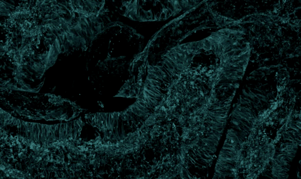
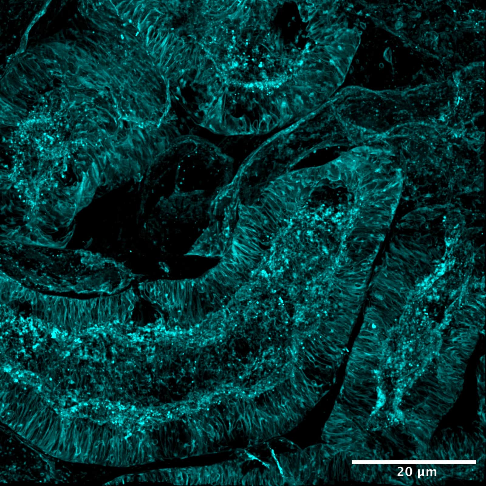
Here is shown a 3D volume imaging of a 16 µm mouse kidney tissue section stained with Alexa Fluor 488 wheat germ agglutinin (WGA) and acquired with CrestOptics Video Confocal Super-resolution system (VCS). WGA is one of the most widely used lectins in cell biology and in mouse kidney labels elements of the glomeruli and convoluted renal tubules. Observing a tissue represents a useful tool for demonstrating microscope optical sectioning properties.