Please, tell us about your scientific career and your experience in microscopy.
I come from a scientific background that is a mix between chemistry and physics which culminated into a PhD in Nano-scale material science. Then, during my post-doc I had the chance to keep doing a lot of different kinds of imaging, such as light microscopy, scanning probe microscopy, and even approach to electron microscopy. Imaging has always been a central part of my scientific interest and all projects I carried out involved a big component of scientific imaging development. During that time, I figured out that my interests were more in the technical, engineering side of imaging which led me to look for a job with a relevant component of that. A core facility is the ideal place to be exposed to many different imaging techniques on a daily basis but also for me to face the challenge of being involved in life science, learning every day from the application side of the research.
“Imaging has always been a central part of my scientific interest.”
Which is your current role at the D-BBSE Single Cell Facility at the ETH Zurich and how the facility is organized?
Here, at the Single Cell Facility led by Dr. Thomas Horn and located in Basel, I take care of the day-to-day microscopy tasks on our different setups, ranging from training and assisted acquisitions to maintenance, QC and customization. At the Department of Biosystems Science and Engineering” we have a wide variety of application-oriented research, also because of the many collaborations with local partners in the pharmaceutical industry. Although everything is oriented around novel biology, the experimental approach can vary from single cells and subcellular processes to large organoids or tissue explants. This variety is what makes our job very interesting and challenging at the same time, since the facility must cover different nuances in how you optimize your imaging experiment.
The general philosophy in our facility is, if possible, to train users to be imaging aware and become autonomous in optimizing their imaging experiments. In short, the goal is to have everyone understand what choices they can make, and how it will affect their imaging and biology. Reaching this level of maturity is usually a (iterative) process, and in some cases not feasible due to limited time-frame available to the trainee. In such cases, we try to optimize the process in a way that is the most time efficient for both users and us. Within the facility we have several staff of complementary backgrounds, with microscopy as the overlapping aspect. This way, we have expertise in-house on all facets of imaging ranging from cell biology (e.g., sample preparation) to optics and engineering (e.g., acquisitions, customizations) to image processing and analysis. Obviously, all aspects are interconnected, and with such a mixed group we all continuously learn a lot from each other as well.
“The complementarity of our staff expertise is important since our users have the same diverse backgrounds.”
Another important aspect we emphasize in our facility is matching the appropriate microscopy modality to the experimental question at hand. The typical test–learn–improve experimental feedback is faster if you start from simpler widefield acquisition and only move to more complex or high-end microscopy modalities as the scientific project and goal matures.
“We teach our users to appropriately escalate the complexity of imaging from widefield to super-resolution as the project matures”
How did you end up buying CrestOptics X-Light V3 spinning disk, and which are the applications that are taking advantages of it?
About one and a half year ago, we decided to expand our inverted spinning disk capacity after assessing our user base needs. It turned out that we needed more high-throughput confocal capacity and a system optimizable for long term experiments. At that time, we were simply limited by the number of experiment slots we could offer, especially to multi-day live cell experiments looking at cell proliferation, cell fates or signalling dynamics.
In addition to that, we experienced in our department, but also in general in life science, a growing demand to get more data from experiments, with more screening type approaches to imaging. A way to fulfil these requests is to have a flexible and fast system with a large field of view, which is exactly the type of features that the CrestOptics X-Light V3 offers. When we had the chance to test the CrestOptics X-Light V3, we were very happy with how it performs; with the larger FOV we can capture a lot more of our samples in the same time frame, and it allows us to do many things we could not do somewhere else (e.g., easily switching from widefield to confocal).
“The main goal we wanted to reach was to have a high-throughput spinning disk system and we largely met our needs with the CrestOptics X-Light V3.”
Which are the applications that are taking advantages of the X-Light V3 spinning disk?
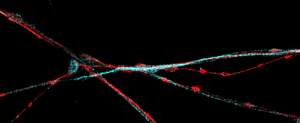
Some of the main imaging applications we plan to run on the CrestOptics system are those multiplexing spatially resolved fluorescence readouts in an iterative, sequential approach (e.g., 4i or seqFISH protocols). Our initial tests on the CrestOptics spinning disk worked very well and we could also benefit from its easy switching between widefield and confocal mode. In addition to that, we are extensively making use of the JOBS scripting features included in the NIS-Elements software (by Nikon) to automate lots of processes. For instance, we incorporated the required pumps and multi-fluidics peripherals within our imaging scripts to design the experiment and minimize the external influences during the acquisition process
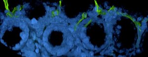
Of note, the introduction of the emission iris in the spinning disk is a special feature of the CrestOptics X-Light V3 and we have found it quite useful, especially for applications where the sample is intrinsically scatter-prone, such as live cells growing on a membrane. By closing the emission iris to the suggested position or even beyond, the reduction of scattering background is clearly evident and hence a significant improvement of the imaging quality can be obtained.
Could you please describe the microscope set-up equipped with CrestOptics X-Light V3?
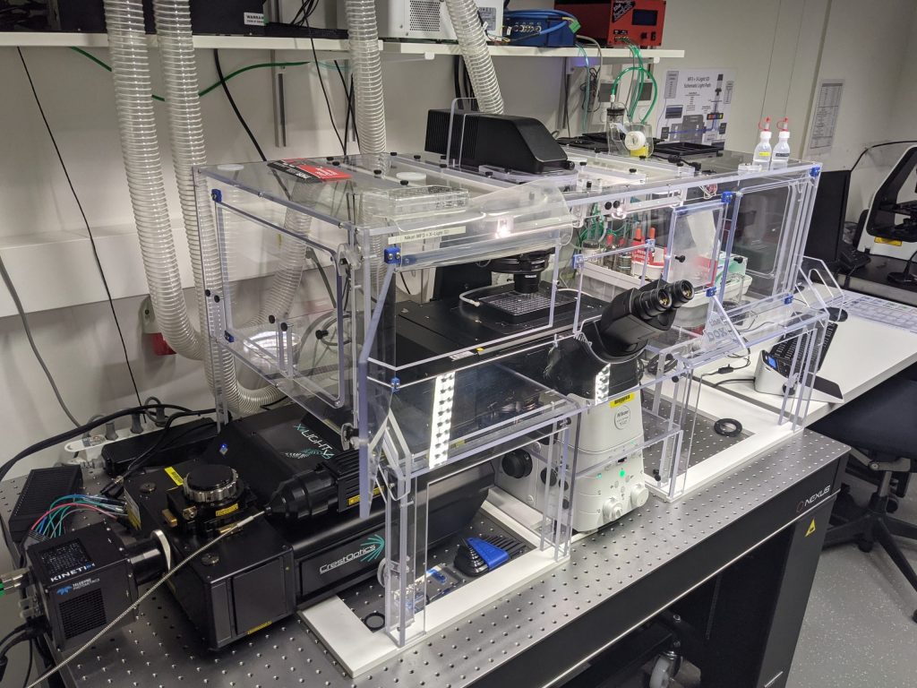
We currently have an inverted Nikon Ti2 microscope equipped with CrestOptics X-Light V3, Kinetix camera (Photometrics) and Celesta (Lumencor) laser source. To allow long-term experiments we also have a large temperature-controlled incubator box around the entire system, with an additional smaller chamber around the sample connected to CO2 and humidity controls.
If we compare CrestOptics spinning disk with standard confocal point scanning, we can go easily much faster with the spinning disk, such that we can image up to 10 times more view fields, compared to point scanning for the same time interval, with less invasive exposure.
“With CrestOptics X-Light V3 we can accentuate either for high speed, or more field of view, or for number of images per time interval.”
How do you see the future of fluorescence microscopy to keep up with innovative biological applications and researcher requests?
I look at microscopy from a facility point of view, so everything that can be modular and easily applicable to a wide range of applications it will always be of interest for situations like ours. What we see as a facility is that things are moving towards faster and bigger approaches where you do more screening, bigger field of view, more things in a short amount of time because of technological advancements and therefore you can get a larger data set and a better overview to answer your biological question. In conclusion, within this scenario the spinning disk and its evolutions will play a central role.